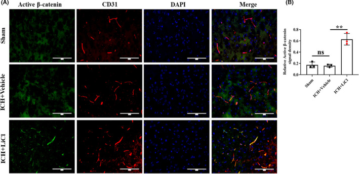
FIGURE 4.

Lithium upregulated Wnt/β‐catenin signaling in the brain endothelium after ICH in vivo. (A) Coimmunofluorescence staining for active β‐catenin (green) and CD31 (red) around the hematoma areas. Scale bar, 100 μm. (B) The active β‐catenin signal density was normalized to the CD31 signal area. n = 3 mice per group. **p < 0.01. ICH, intracerebral hemorrhage
