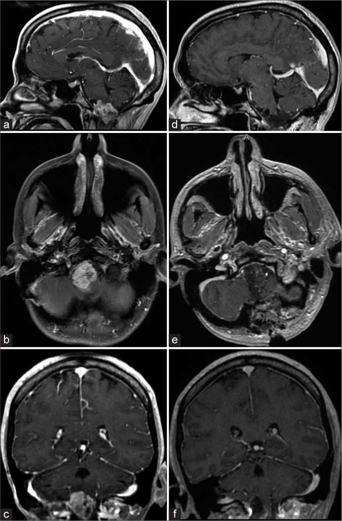
Figure 1:

MRI of astroblastoma MN1-altered tumor. (a-c) Sagittal, axial, and coronal preoperative T1 MRI revealing an intra-axial, heterogeneously enhancing lesion centered in the cervicomedullary junction with rostral extension into the pontomedullary junction and caudal extension into the C1-C2 spinal cord. (d-f) Sagittal, axial, and coronal postoperative T1 MRI demonstrating successful resection of the lesion.
