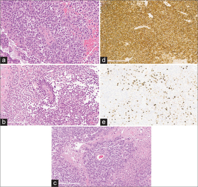
Figure 2:

Histopathology of astroblastoma MN1-altered tumor. (a-c) Hematoxylin and eosin staining exhibiting epithelioid neoplasm with highly mitotic tumor cells with an average Ki67 proliferation index of 19%, distinct cell borders, variable sheet-like, pseudopapillary, and trabecular architecture, and multiple foci of bland necrosis with hyalinized vessels. (d) Positive for EMA immunoreactivity, (e) positive for Ki67 staining (average index of 19%, hotspots of 26%).
