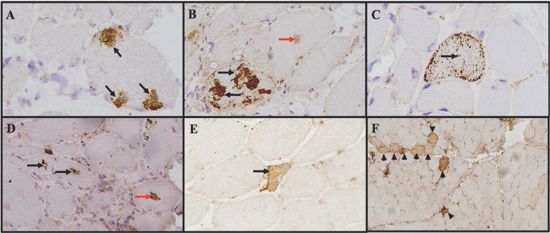
Fig. 1.

Examples of different patterns of p62 immunostaining in patients with myositis.
A) subsarcolemmal aggregates, B) sarcoplasmic aggregates, C) punctate pattern, D) perivacuolar pattern, E) necrotic fibre and F) perifascicular pattern. Red arrows indicated fine grouped aggregates.
