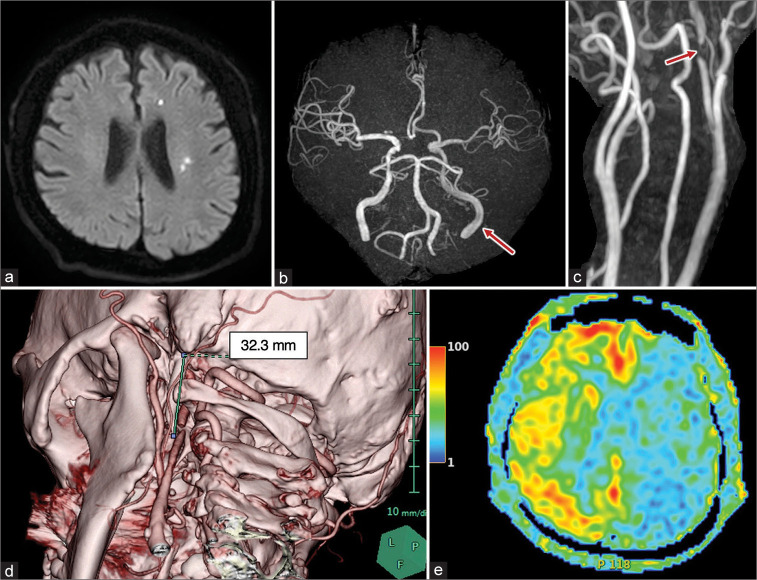
Figure 1:
Radiological images at initial symptom onset are presented. DWI showed multiple hyperintense lesions at the left hemisphere (a) and intracranial blood flow was impaired at the lesion side on magnetic resonance angiography (b: Red arrow). The blood flow signal was absent at the high cervical portion of the ICA (c: Red arrow). 3D-CTA revealed an enlarged styloid process, whose length was 32.3 mm (d), and a cerebral blood flow study by arterial spin labeling showed a remarkable CBF decrease at the affected side (e).

