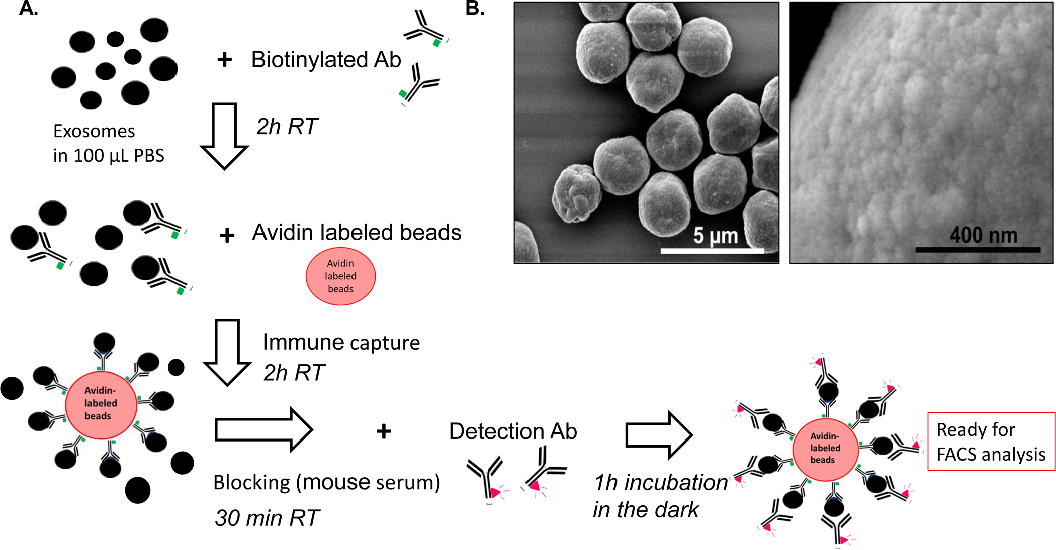
Figure 1: Immune capture and detection of the exosome cargo by on-bead flow cytometry.

A: A schema for the capture of exosomes onto beads and antigen detection on exosomes. After exosome isolation from plasma via SEC, immune capture with a biotinylated Ab (e.g., anti- CD63 Ab) is followed by incubation with streptavidin beads. For antigen detection, exosome/Ab/bead complexes are incubated with fluorochrome-conjugated detection Ab, and MFI of the targeted antigen is determined by on-bead flow cytometry. B: Visualization of exosomes captured on beads via scanning electron microscopy (SEM). Note an excess of exosomes that are packed on beads. Titrations of exosomes and beads are necessary to avoid excessive exosome “crowding” on the bead surface.
