
Figure 7. On-bead flow cytometry for exosomes disrupted with 0.3% Triton X.
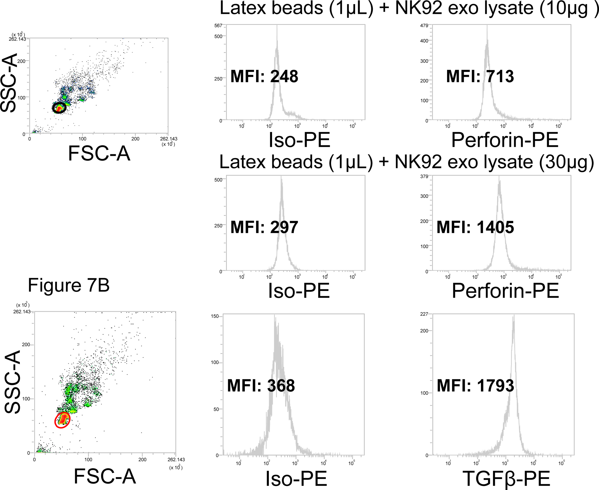
Exosomes produced by NK92 cells were treated with 0.3% Triton X-100 in PBS as described in Methods. Lysates were placed on latex beads (1μL) at the pre-determined exosome/beads ratios for detection with PE-labeled anti-perforin Abs. Upper panels: flow cytometry for perforin in 10μg lysate of exosomes produced by NK92 cells. Lower panels: flow cytometry for perforin in 30μg 30ug of the same lysate. At 50μg of lysate, MFI did not increase (not shown), indicating that saturation was reached at 30μg. In B, detection of intraluminal TGF-β on exosomes produced by NK92. We have previously reported that these exosomes carry TGF- β based on Western blotting of exosomes (25). In A and B, note that with latex beads, the gate should be carefully set to exclude bead aggregates. .