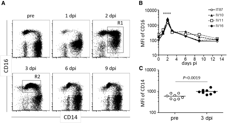
FIG. 4.
Altered CD14 and CD16 expression on myeloid lineage cell populations after CHIKV infection. The CD45+CD3−CD20−CD8− population (as described in Fig. 1) was examined for CD14 and CD16 expression by using serially collected blood after CHIKV infection. (A) Representative flow cytometric profiles from one of seven CHIKV-infected macaques are shown. (B) Kinetic of mean fluorescence intensity of CD16 in R1 shown in (A) after CHIKV infection. Statistical difference between day 2 and preinfection time points were analyzed by two-way ANOVA. ****p < 0.0001. (C) CD14 expression in R2 shown in (A) was compared between preinfection and 3 days after infection. The statistical difference was analyzed by Mann–Whitney test.

