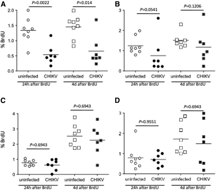
FIG. 7.
Lymphocyte proliferation in acute CHIKV infection. CHIKV-infected macaques were administered BrdU at 2 days postinfection. Blood was collected serially after BrdU administration and stained with anti-CD3, CD4, CD8, CD20 and NKG2a, and BrdU antibodies for flow cytometric analysis. The data were compared to those form uninfected macaques. The cellular proliferation of CD4+ T cells (CD3+CD20−CD8−), CD8+ T cells (CD3+CD4-CD8+), CD20+ B cells (CD3−CD20+), and NK cells (CD3−CD20− CD8+NKG2a+) is shown in (A–D), respectively. The statistical differences between uninfected and CHIKV-infected groups were calculated by Mann–Whitney test.

