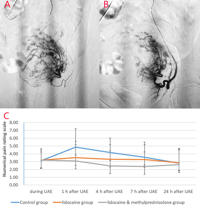
Figure 3.
Images from a patient in the study group and a patient in the lidocaine group. (A) Before lidocaine injection and (B) after lidocaine injection. Left uterine arteriography images show uterine artery spasm after lidocaine injection, as indicated by the white arrow. (C) Pain score curve of the three groups.

