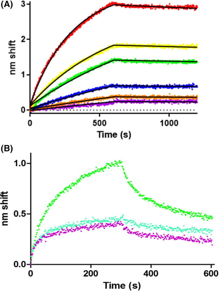
Fig. 2.

JNK1α1 binds to NOS3 in a concentration‐dependent manner. (A) BLI sensorgram of JNK1α1 binding to NOS3. Purified NOS3 at concentrations ranging from 25 to 800 nm (purple, 25 nm; orange, 50 nm; blue, 100 nm; green, 200 nm; yellow, 400 nm; red, 800 nm) were assayed for binding to ligand GST‐JNK1α1. Raw data are shown with fits to a one‐state association‐then‐dissociation model. The association phase was from 0 to 600 s, whereas dissociation was from 600‐1200s. (B) No evidence of JNK1α1 binding to NOS1 beyond nonspecific binding. Green denotes response to ligand calmodulin, blue represents response to ligand JNK1α1 and purple is no ligand (NS) control.
