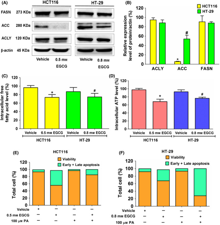
Fig. 2.

The effect of EGCG on expression of enzymes in the DNL pathway, free fatty acid and ATP levels in colorectal cancer cells, HCT116 and HT‐29 cells. Cells were incubated with EGCG at an IC50 concentration for 24 h. (A) An immunoblotting assay was used to assess the expression of enzymes in the DNL pathway. (B) A histogram depicts relative protein expression in comparison to β‐actin. (C) The histogram depicts free fatty acid levels and (D) ATP levels compared to the vehicle control. Apoptotic assay in (E) HCT116 and (F) HT‐29 cells using annexin V/PI staining in EGCG, palmitate (PA) at 100 μm, and a combination of EGCG and PA. Data are expressed as the mean ± SD from at least a triplicate of n = 3. *P < 0.05 and # P < 0.05 compared to the control of each cell. All data were analyzed using one‐way ANOVA with Tukey’s post‐hoc test.
