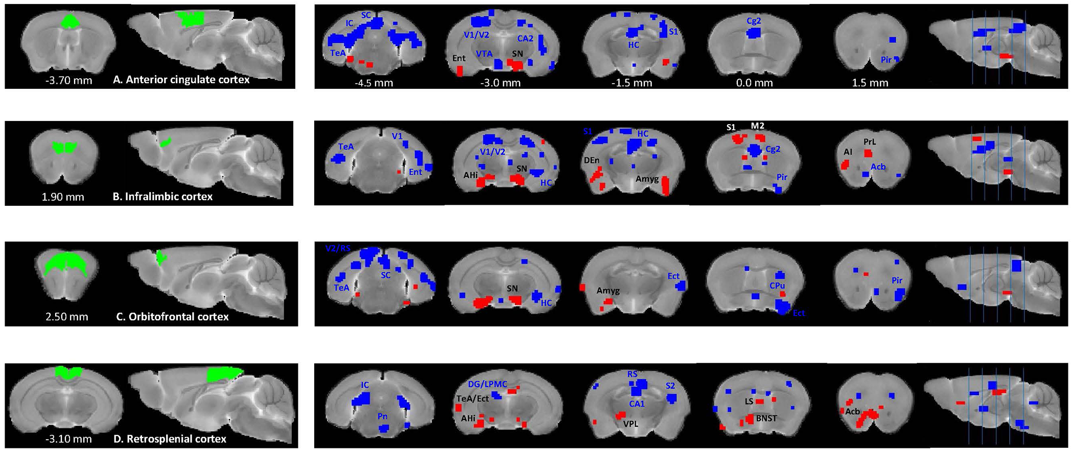
Figure 4. Between-group comparisons of seed-based rs-FC for the cortical brain regions between Df(h22q11)/+ and wild type mice.

Two-sample t-tests (p <0.01) show significantly higher (red) and lower (blue) connectivity between anterior cingulate cortex (A), infralimbic cortex (B), orbitofrontal cortex (C) and retrosplenial cortex (D) and multiple cortical and subcortical brain regions in Df(h22q11)/+ mice, compared to the control group. Coordinates are in mm to Bregma. Abbreviations: AHi – amygdalohippocampal area, Amyg - amygdala, Ect – ectorhinal cortex, LPMC – lateral posterior thalamic nucleus mediocaudal part, Pn – pontine nuclei, V1 – primary visual cortex, VPL – ventral posterolateral thalamic nucleus. Other abbreviations are the same as in Figure 3.
