Figure 21:

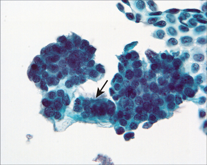
Three-dimensional cell groups are present, associated with a sheet of mesothelial cells. Some of the cells showed cilia (arrow). (Modified PAP stain, 40X.)

Three-dimensional cell groups are present, associated with a sheet of mesothelial cells. Some of the cells showed cilia (arrow). (Modified PAP stain, 40X.)