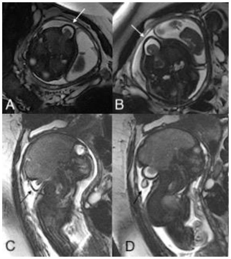
Figure 2.
An in-utero male fetus with occipital and parietal encephalocele
Findings: Intra uterine fetal MRI showing occipital encephalocele with herniation of meninges and brain parenchyma through the bone defect in axial (A, B) and sagittal plane (C, D).
Technique: Intra-uterine fetal MRI with non-contrast T2 weighted images (1.5 Tesla, TR 4000, TE 90) in axial (A, B) and coronal (C, D) planes.

