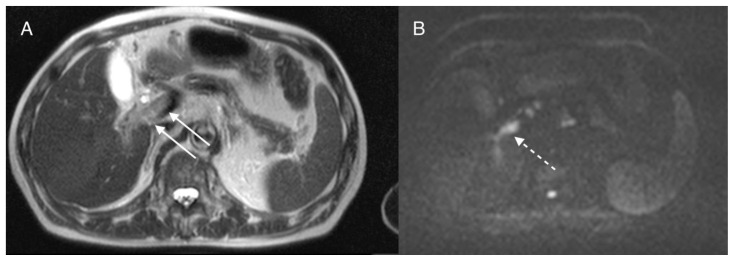
Figure 10.
70-year-old man with pylephlebitis
FINDINGS: Axial MRI reveals absence of normal flow void on T2WI (A arrows) and hyperintensity on DWI within the portal vein (B dashed arrow).
TECHNIQUE: Axial T2WI was performed using a SIEMENS Symphony 1.5T MRI scanner with a TE: 108, TR: 950, axial DWI was performed with a TE: 76, TR: 5300, B-value: 800.

