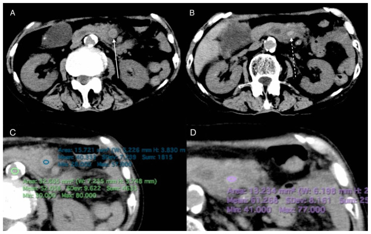
Figure 2.
82-year-old woman with pylephlebitis
FINDINGS: Axial NECT shows intravascular hypo- (A arrow) and hyper-attenuation (B dashed arrow) in the peripheral and central SMV, respectively. The intravascular hypo- and hyper-attenuation, and the aorta show 40.3, 61.3, and 52.1 HU, respectively (C,D).
TECHNIQUE: Non-enhanced axial CT images were acquired with TOSHIBA Aquilion 16-slices CT scanner at 93 mAs, 120 kV, and 8 mm slice thickness.

