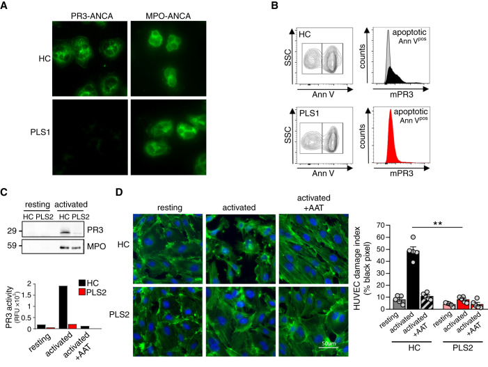
Figure 2.
PLS neutrophils produce a negative clinical PR3-ANCA immunofluorescence test, expose less PR3 on apoptotic neutrophils, and cause less NSP-mediated endothelial injury. (A) Indirect immunofluorescence using HC and PLS neutrophils and sera from patients on PR3 and MPO, respectively. (B) A percentage of overnight cultured neutrophils showed constitutive apoptosis by Annexin V staining. mPR3 was analyzed after gating on apoptotic (Annpos) cells. (C) cf-SN from resting and activated (2.5 µM ionophore A23187) HC and PLS neutrophils were assessed for PR3 protein by immunoblotting and proteolytic activity by FRET assay. (D) Confluent HUVEC monolayers were incubated with cf-SN from resting and activated HC and PLS neutrophils, respectively. When indicated cf-SN from activated neutrophils were treated with α1-antitrypsin (AAT) before EC incubation. EC damage was visualized by actin staining with phalloidin-FITC (green) and nuclear DAPI staining (blue), and quantified by determining the black pixel areas using a Leica microscope (×40) and ImageJ 1.48v software. ** P<0.01.

