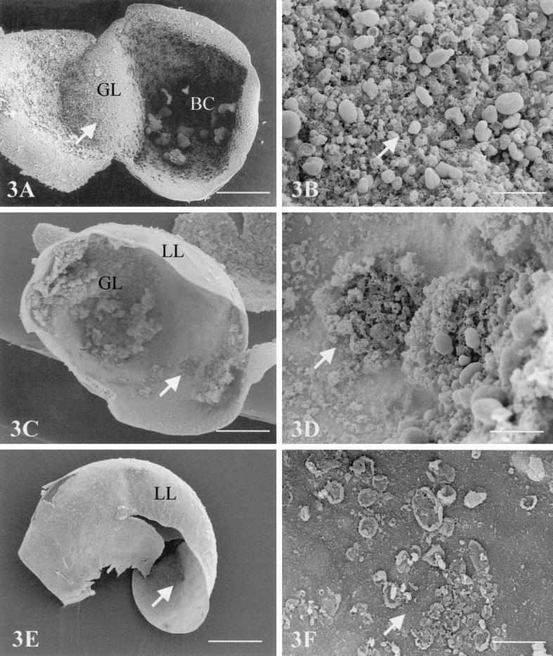
FIG. 3.
SEM nontreated (A and B) or ABZSO-treated (C to F) E. multilocularis metacestodes. GL, germinal layer; LL, laminated layer. (A and B) Control metacestodes cultured in vitro in the presence of DMSO (1:1,000) but in the absence of any drugs. Note that most cells exhibit an intact morphology. Bar = 1.2 mm (A) or 200 μm (B). (C to F) SEM of metacestodes cultured in vitro in the presence of ABZSO for 10 days (C and D) and of ABZSN for 14 days (E and F). (C) Large portions of the germinal layer have disintegrated after 10 days of drug treatment and are detached from the laminated layer (bar = 900 μm). (D) Higher-magnification view of image in panel A (bar = 200 μm). (E) After 14 days, only metacestode “ghosts,” comprised of the acellular laminated layer, are found (bar = 1.2 mm). (F) Higher-magnification view onto the interior surface of the laminated layer. Note the presence of largely destroyed cells (bar = 180 μm).

