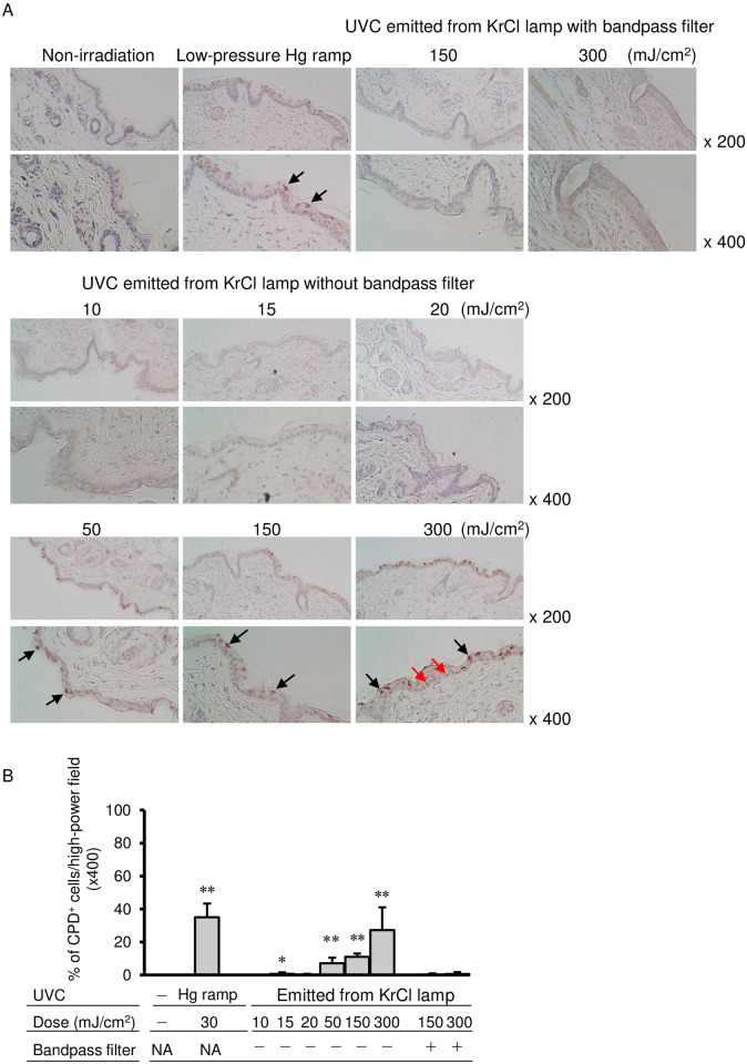
Fig 2. Histological analysis of dorsal skin of mice irradiated with UVC emitted from a KrCl lamp with or without a bandpass filter.
The dorsal skin of mice was irradiated with UV radiation from a low-pressure Hg lamp, UVC emitted from a KrCl lamp with a bandpass filter at 150 and 300 mJ/cm2 or UVC emitted from a KrCl lamp without a bandpass filter at 10, 15, 20, 50, 150 and 300 mJ/cm2. The skin specimens were stained with anti-CPD antibody as described in the Materials and Methods section. Black arrows indicate CPD-positive cells, and red arrows indicate CPD-positive cells in the basal layer of the epidermis (A). CPD-positive and CPD-negative cells were quantified by counting cells in dermis in four random high-power fields (x400) for each preparation, and percent of CPD-positive cells was determined. Data are presented as the mean ± standard deviation. The non-irradiated group and each irradiated group were statistically compared. ** P < 0.01, * P < 0.05 (B). CPD: cyclobutyl pyrimidine dimers; Hg: mercury; KrCl: krypton chloride; NA: not applicable; UVC: ultraviolet C.

