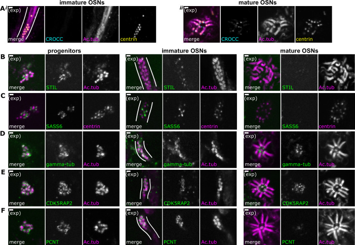
Figure 3. Composition of centriole groups during olfactory sensory neuron (OSN) differentiation.
(A) The cohesion fiber and striated rootlet protein rootletin is absent during centriole migration but is gained at the mature dendritic knob. Expansion microscopy, maximum z-projection of confocal stack. In all side-view images, apical is oriented toward the top of the image. Cyan: staining for Rootletin (CROCC). Magenta: staining for acetylated tubulin; yellow: staining for centrin. (i) Side view of migrating centrioles. Centrioles below the apical surface, in the dendrite of an immature OSN. (ii) Centrioles at the apical surface in the same sample as those shown in (Ai). Scale bars = 2 μm. (B–F) Expansion microscopy – single-plane fluorescence images of centrioles in progenitor cells (left column), immature OSNs with migrating centrioles (middle column, white lines outline an OSN dendrite), and mature OSNs imaged en face (right column). Scale bars = 2 μm. (B) The immature centriole protein STIL is present in progenitors and immature OSNs. Green: staining for STIL; magenta: staining for acetylated tubulin. (C) The immature centriole protein SASS6 is present in progenitors and immature OSNs. Green: staining for SASS6; magenta: staining for centrin. (D) The pericentriolar material protein gamma-tubulin is present throughout OSN differentiation. Green: staining for gamma-tubulin; magenta: staining for acetylated tubulin. (E) The pericentriolar material protein CDK5RAP2 is present throughout OSN differentiation. Green: staining for CDK5RAP2; magenta: staining for acetylated tubulin. (F) The pericentriolar material protein pericentrin (PCNT) is present in progenitors and immature OSNs. Green: staining for pericentrin (PCNT); magenta: staining for acetylated tubulin.
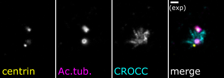
Figure 3—figure supplement 1. Expansion microscopy staining of rootletin in cycling cells.


