. 2019 May 21;9(28):15887–15899. doi: 10.1039/c9ra02368b

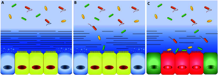
Fig. 2. (A) Schematic representation of intestinal epithelium comprising epithelial cells (yellow), mucus-secreting goblet cells (blue) and two well-defined mucus layers. The mucus has a double-layer structure: the first layer firmly attached to the mucosa (dark-blue with lines) and a loose outer layer (represented by the light-blue without lines in the top background), which may be removed easily. The thickness ratio of the attached and loose layers increases along the intestinal tract. In healthy conditions, bacteria grow only in the loose layer. (B) When the attached layer is defective or degraded by pathogens (changes in the blue shades and number of lines), epithelial cells are in direct contact with bacteria. (C) As a consequence, serious diseases such as colitis and chronic inflammation can occur (green and red colours were used to indicate a generic pathological cellular conditions).

Fig. 2