. 2019 May 31;9(30):16929–16947. doi: 10.1039/c9ra01253b

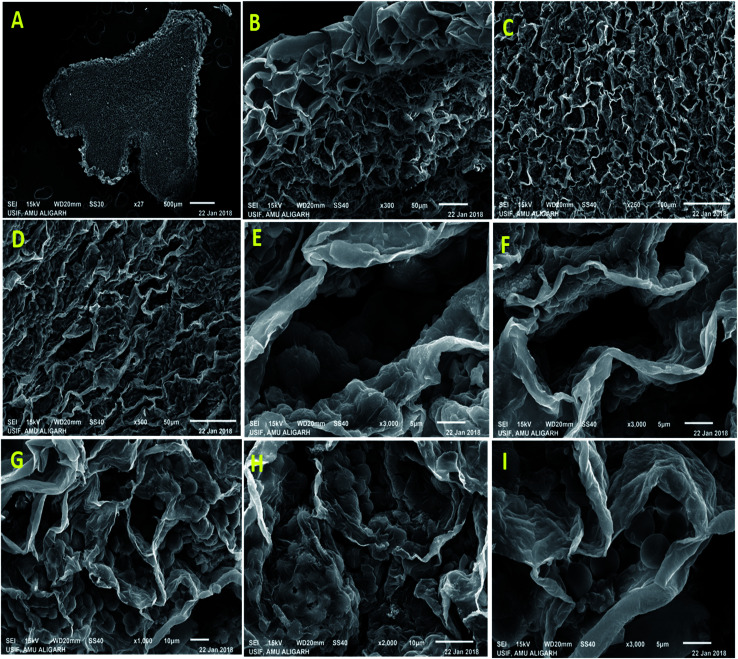
Fig. 9. A low magnification scanning electron micrograph of P. sativum nodules cut longitudinally through its long axis showing overall nodule structure. A portion of nodule scanned from inside to the surface showing (A) central tissue (B) cortex tissue (C) empty cortex tissue and (D) collapsed empty cortex tissue which make up the outermost surface layer of the nodule, (E) showing the structure of infected cells packed with healthy Y and T shaped bacteroides in untreated nodules and (F) depicts the empty cells representing the absences of bacteroids, and (G–I) displayed the infected cells containing deteriorating bacteroides with zoogloeal mass covered with the thin layers.

Fig. 9