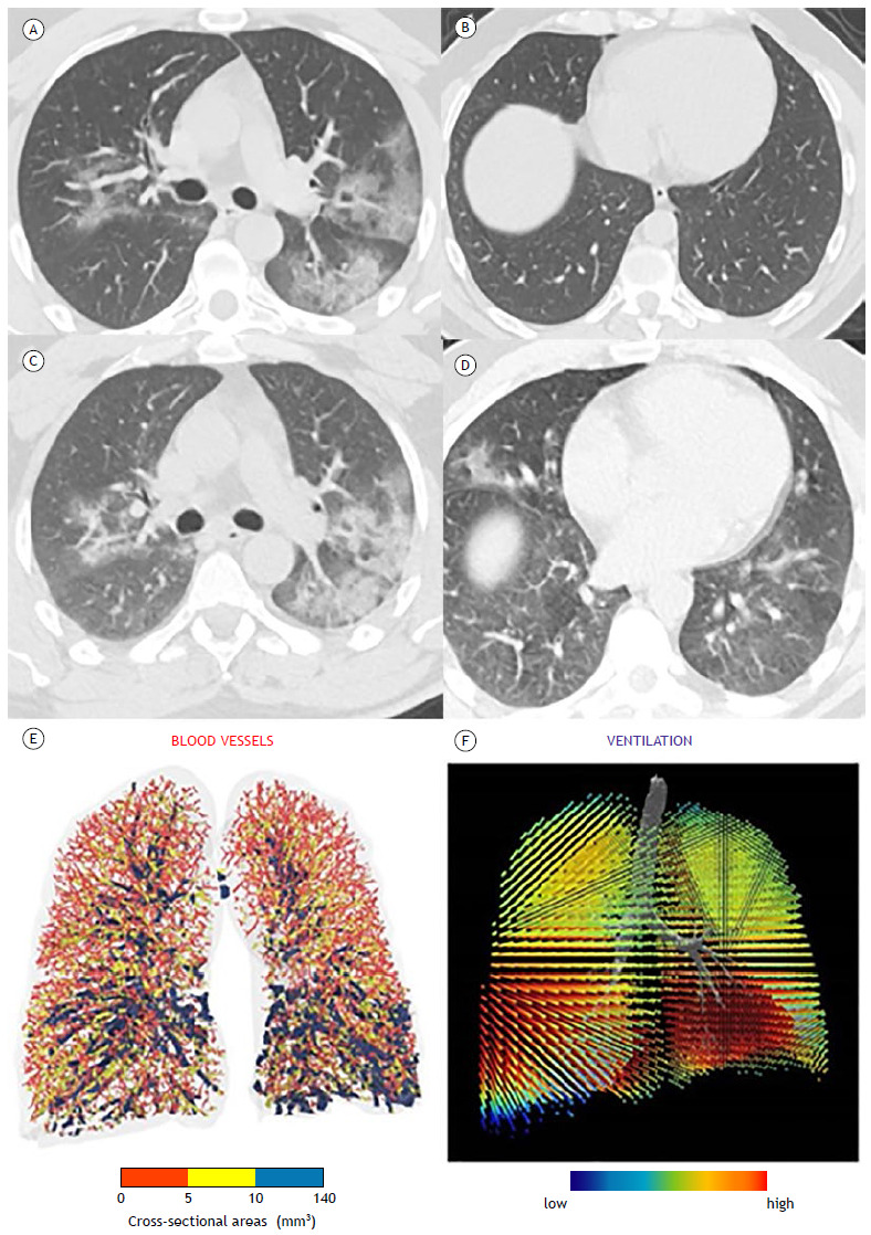
Figure 2. Typical appearance of COVID-19-25-49% parenchymal involvement. HRCT axial images obtained during inspiratory (in A and B) and expiratory (in C and D) acquisitions showing air trapping. In E, three-dimensional visual representation of blood vessels colored according to their size (red, yellow, and blue corresponding to small, mid-sized, and large vessels, respectively). Cross-sectional areas < 5 mm3 are sparse throughout the lung, indicating severe diffuse vasoconstriction even in areas without consolidation. In the ventilation map (in F), most areas of the lung are colored red, representing a normal expansion of lobar volumes between inspiration and expiration, even in areas where there is severe vasoconstriction of small blood vessels.