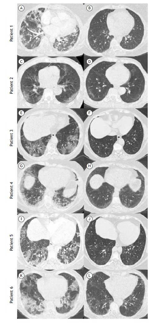
Figure 1. Chest CT scans of the patients studied. Patient 1: in A, a scan during the acute phase showing bilateral ground-glass opacities (GGO), consolidations, and parenchymal bands; in B, a scan after 15 months of follow-up showing subtle peripheral and posterior GGO. Patient 2: in C, a scan during the acute phase showing bilateral and peripheral GGO; in D, a scan after 7 months of follow-up showing subtle GGO with subpleural curvilinear lines and small dilated bronchioles in the right lower lobe. Patient 3: in E, a scan during the acute phase showing bilateral GGO and crazy-paving pattern; in F, a scan after 6 months of follow-up showing subtle scattered GGO. Patient 4: a scan during the acute phase showing bilateral and peripheral GGO and consolidations; in H, a scan after 4 months of follow-up showing subtle bilateral and peripheral GGO. Patient 5: in I, a scan during the acute phase showing bilateral GGO; in J, a scan after 10 months of follow-up showing subtle GGO and mosaic attenuation in the lung parenchyma. Patient 6: in K, a scan during the acute phase showing bilateral GGO and consolidations; in L, a scan after 7 months of follow-up showing bilateral GGO with some dilated bronchioles.

