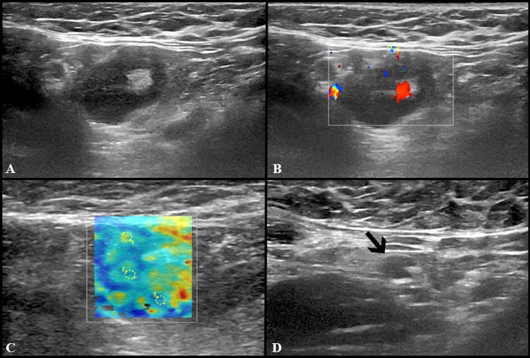
Fig. 3.
Additional images from the same subject reported in Fig. 2: an enlarged axillary lymph node (17 × 11 mm) with fatty hilum but diffuse cortical thickening (10 mm) and hilar vascularization (A and B) was detected. Shear wave elastography showed soft-intermediate consistence (C: k1 = 35 kPa, k2 = 17 kPa and k3 = 13 kPa). At the follow-up US scan (D) performed after 4 weeks lymph node size reduction and normalization of cortical thickness were observed (black arrow)

