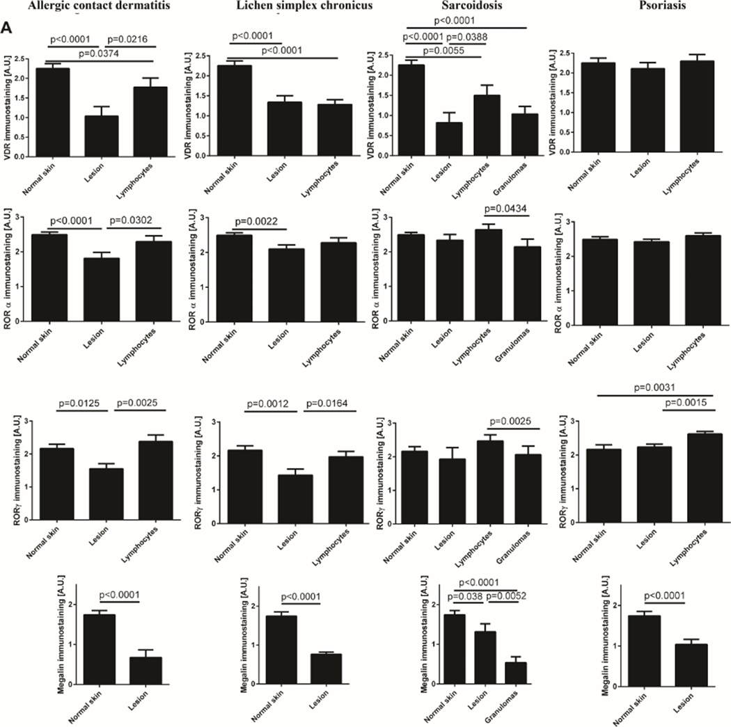
Figure 2.
Expression levels of VDR, RORα and RORγ and LRP2/megalin. A) The expression measured by immunohistochemistry in psoriasis, allergic contact dermatitis, lichen simplex chronicus and sarcoidosis. Statistically significant differences are denoted with p values as determined by Student’s t-test. B). The expression measured by RNA-seq in psoriasis and normal skin (https://www.ebi.ac.uk/gxa/home; 40).


