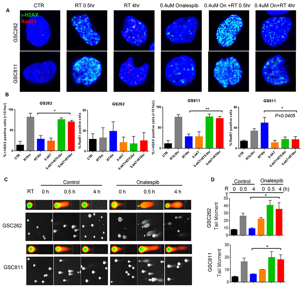
Figure 3.

Effect of onalespib on IR-induced DNA damage and subsequent repair in GSC811 and GSC262 cells. (A) Immunofluorescence images (upper panels) of time-course of appearance and resolution of γ-H2AX (marker of DNA damage) and RAD51 foci (marker of ongoing HR repair) after IR-induced DNA damage alone or in combination with onalespib in GSC (upper panels). (B) Quantitation of immunofluorescent γ-H2AX foci and Rad-51 foci in GSCs treated with IR alone or IR+onalespib. Graph represents at least three independent experiments (*=p<0.05, **=p<0.005, two-tailed p test, error bars=SEM). (C) Comet assay imaging of GSC811 and GSC262 cells at 0.5h or 4h after treatment with IR alone or IR+onalespib (0.4μM) showing persistence of comet tails in the onalespib treated cells at 4h post-treatment indicating failure to repair IR-induced DNA strand breaks. (D) Quantification of comet tail moment indicating onalespib-mediated abrogation of cellular DNA repair after IR. Graphs represent triplicate experiments (*=p< 0.05). (RT=radiation therapy; AT: onalespib)
