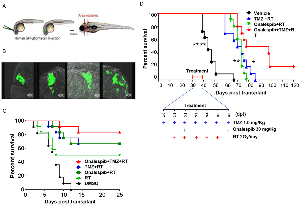
Figure 6.

Onalespib-mediated effects on chemoRT therapy and effects on survival in a zebrafish and nude mouse intracranial glioma xenograft models. (A) Schematic diagram of intracranial human U251HF-GFP glioma-xenotransplanted zebrafish model. (B) GFP-expressing diffusely infiltrative tumor cells in intracranial glioma xenograft seen in transverse sections at various levels in the zebrafish brain. (C) Kaplan-Meier survival curves for zebrafish glioma (n=11-12 animals/group). U251HF-GFP xenotransplanted zebrafishes treated with DMSO, RT (2 Gy/day for 5 days), TMZ (10 μmol/L) plus RT, onalespib (0.5 μmol/L) plus RT, or a combination of TMZ (10 μmol/L) and onalespib (0.5 μmol/L) with RT. (D) Kaplan-Meier survival curves for U251HF-Luc xenotransplanted nude mice (n=10-12 mice/group) treated with vehicle (PBS), TMZ (1.0 mg/Kg) + RT (2 Gy/day for 5 days), onalespib (20 mg/Kg) + RT, or a combination of onalespib and TMZ+RT; differences in survival were assessed by the log-rank test. (combination vs. vehicle, ****, P < 0.0001; combination vs. onalespib + RT, **, P < 0.01; combination vs. TMZ + RT, *, P < 0.05) (TMZ=temozolomide; RT= radiotherapy).
