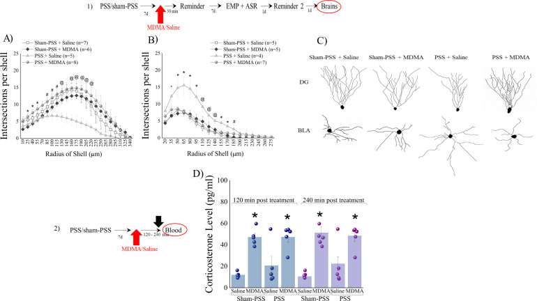
Fig. 2. MDMA treatment paired with a trauma-cue at 7 days after trauma, normalized morphological indicators.
The top panel (1) depicts the experimental protocol. Vertical arrows represent intraperitoneal MDMA (5 mg/kg) or saline injection. Rats were exposed for 10 min to predator-scent stress (PSS) or to sham-PSS on day 0. On day 7, rats received MDMA (sham-PSS + MDMA: n = 10; PSS-exposed + MDMA: n = 10) or saline (sham-PSS + saline: n = 11; PSS-exposed + saline: n = 10) and 30 min later were exposed to a trauma-cue for 10 min. On Day 16, rats were sacrificed and their brains collected for morphological staining. A Sholl analysis for intersections per 15-μm radial unit distance of dentate gyrus granule cells from the suprapyramidal blade. * Sham-PSS + MDMA ≠ PSS + MDMA, p < 0.05. #PSS + saline ≠ PSS + MDMA, p < 0.05. @PSS + saline ≠ PSS + MDMA, sham-PSS + saline, p < 0.05. B Sholl analysis for intersections per 15-μm radial unit distance of pyramidal neurons of the basolateral amygdala. ^PSS + saline ≠ sham-PSS + saline & sham-PSS + MDMA, p < 0.03. * PSS + saline ≠ sham-PSS + saline & sham-PSS + MDMA & PSS + MDMA, p < 0.03. @PSS + saline ≠ sham-PSS + saline & PSS + MDMA, p < 0.05. #PSS + saline ≠ sham-PSS + saline, p < 0.04. C Computer-generated reconstructions of dendritic trees from granule cells and pyramidal cells in all groups. (2) depicts the experimental protocol. Vertical arrows represent intraperitoneal MDMA (5 mg/kg) or saline injection. Corticosterone concentrations (pg/ml) measured at 120- and 240 min post MDMA treatment in sham-PSS treated with saline (Sham-PSS + saline, n = 10) or MDMA (Sham-PSS + MDMA, n = 10), PSS-exposed animals treated with saline (PSS + saline, n = 10), or PSS-exposed treated with MDMA (PSS-MDMA, n = 9). D corticosterone concentrations** (pg/ml) were measured. The experiments described below were performed with two different cohorts of animals; 20 rats were run in one experimental design, and 19 rats in another experimental design. Bars represent group means ± SEM.

