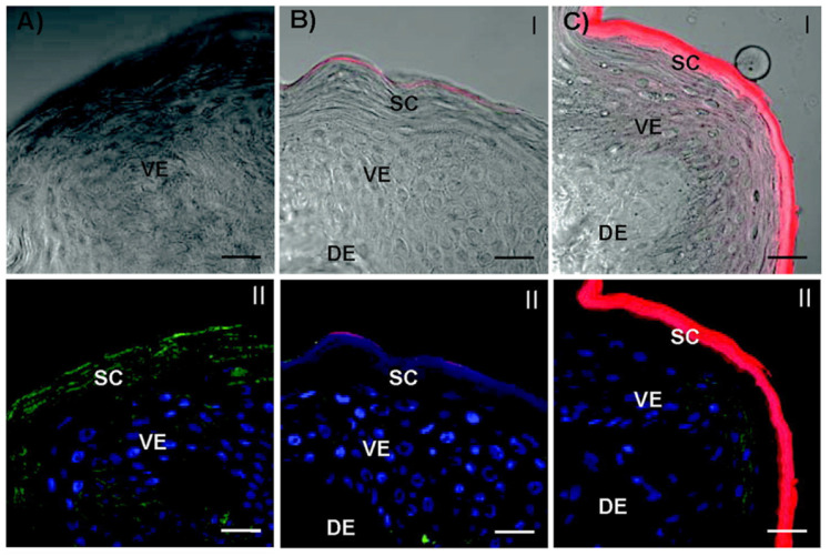
Figure 3.
Confocal laser scanning microscopy images of the skin cross-sections of microtomed porcine skin layers after treatment with dendrimers. (A) Vehicle (ddH2O) control. (B) G4-RITC-NH2. (C) G2-RITC-NH2. (dendrimer conjugates (red), cell membrane stained by WGA-AF488 (green), and nuclei stained by DAPI (blue)). Scale bar: 10 μm. Adapted with permission from reference 48. Copyright 2012 American Chemical Society.

