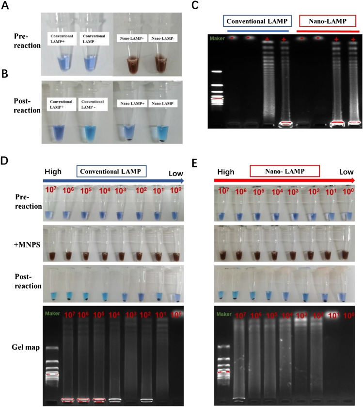
Fig. 3.
A Images of pre-reactions for conventional LAMP (conventional LAMP + and conventional LAMP −) and reactions added MNPs. (B) Post-reaction images for nano-LAMP (nano-LAMP + and nano-LAMP −) after magnetic separation. (C) Gel electrophoresis images of LAMP products in A and B. (D) Color contrast pictures and electrophoretic contrast of HPV DNA diluted to different concentrations (107 copies/mL–101 copies /mL) of HPV-6 DNA were involved in conventional LAMP reactions and in nano-LAMP reactions (E)

