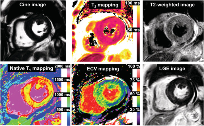
Figure 2.

Baseline cardiovascular magnetic resonance. There was patchy mid‐wall LGE and mildly increased ECV of 36% in the basal septum; no increased global myocardial native T1, T2 values (1334 and 51 ms, respectively) or signal intensities on T2‐weighted images were observed. These findings were inconsistent with the 2018 Lake Louise criteria for a diagnosis of acute myocarditis. (Normal values at our institution: native T1:1314 ± 29 ms, T2:46 ± 5 ms, ECV: 26 ± 5%). ECV, extracellular volume fraction; LGE, late gadolinium enhancement.
