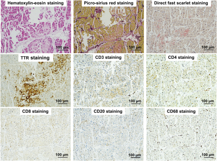
Figure 3.

Pathological findings. Haematoxylin–eosin and picrosirius red‐stained samples showed mild extracellular expansion and minimal lymphocytic infiltrates with mild infiltrative interstitial fibrosis. Anti‐transthyretin immunohistochemistry was positive for transthyretin. There was only a minimal increase in CD3+ T‐lymphocytes, with a predominance of CD4+ T‐lymphocytes, and myocardial CD68+ macrophages, which did not fulfil the pathological diagnostic criteria of myocarditis.
