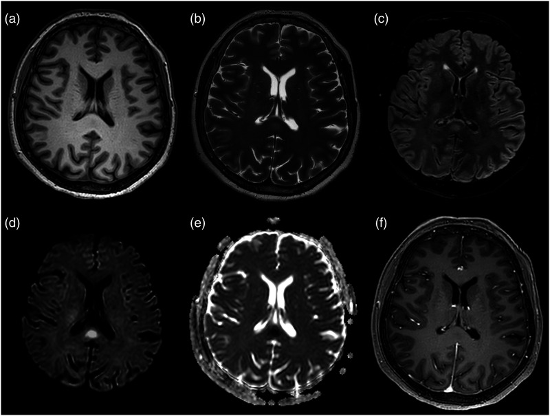
Figure 1.
First axial brain MRI on the 4th day of admission, 8 days after SARS-CoV-2 mRNA vaccine (BNT162b2). (a) T1-weighted images show a mild hypointense oval-shaped lesion in the splenium of the corpus callosum. (b, c) The lesion presents slightly hyperintense signal on T2-weighted and FLAIR images. (d, e) Hyperintense signal on b-2000 diffusion-weighted images and low apparent diffusion coefficient values are noted, consistent with restricted diffusion within the lesion. (f) Contrast-enhanced T1-weighted sequences reveal no contrast enhancement.

