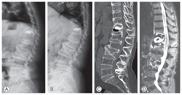
Fig. 2.
Recollapse of a vertebra after percutaneous vertebroplasty in a 73-year-old woman with the intravertebral vacuum cleft sign and instability. Local kyphosis and collapse of the vertebral body are more prominent on a standing lateral radiograph than on a supine radiograph. (A) Lateral standing radiograph. (B) Lateral supine radiograph. (C) A sagittal computed tomography scan shows the intravertebral vacuum cleft sign and recollapse of the vertebral body. (D) A fat-suppressed T2-weighted magnetic resonance image shows high signal intensity around the augmented cement in the vertebral body.

