
Fig. 5.
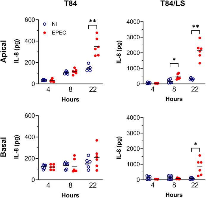
IL-8 secretion in response to EPEC infection by T84 and T84/LS174T epithelia. Cell monolayers were incubated with EPEC or left non-infected (NI) for 4, 8 or 22 h in the VDC. Medium containing gentamicin (50 µg/ml) was added after 4 h to prevent bacterial overgrowth. IL-8 in apical and basal supernatants was quantified by ELISA and expressed as total pg per chamber. Shown are individual data points with means indicated by a line. Data were analysed by two-way ANOVA with Sidak's multiple comparison test, *P<0.05, **P<0.01.