
Fig. 7.
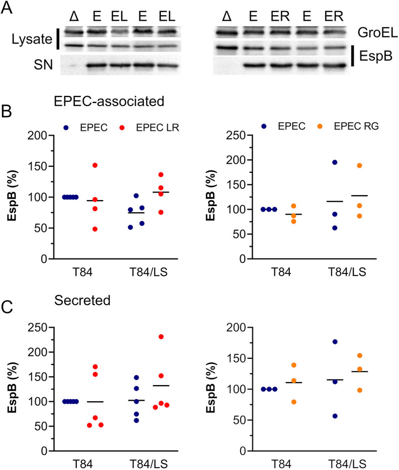
EPEC EspB production and secretion in the presence of L. reuteri and R. gnavus. (A-C) T84 and T84/LS174T epithelia were incubated with EPEC alone (E) or in co-culture with L. reuteri (EL) or R. gnavus (ER) for 4 h. T84/LS174T cells were also inoculated with EPEC ΔescN (Δ) to control for T3S. Apical media were sampled, and bacteria separated from supernatants by centrifugation. Bacterial lysates and concentrated supernatants (SN) were separated by 12% SDS-PAGE, and EspB was detected by western blotting. Membranes were re-probed for GroEL as housekeeping control (A). Bands were quantified by densitometry, and EspB signals were normalized to GroEL controls. EspB levels in EPEC-associated (B) and supernatant (C) fractions are expressed as percentage relative to EPEC-infected T84 cells. Data are plotted as individual data points with means indicated by a line. Statistical analysis was performed by two-way ANOVA with Tukey's multiple comparison test.