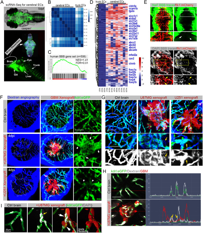
Fig. 3.
The zebrafish GBM xenograft provides a visual readout for the structural and functional changes of the tumor-associated blood–brain barrier (BBB). (A) Schematic showing the strategy of separating cerebral endothelial cells (ECs) from the trunk ECs of zebrafish larvae. (B) Unsupervised clustering analysis based on all significant differentially expressed genes of 12 ECs. (C) Gene set enrichment analysis (GSEA) showing the enrichment of BBB genes in zebrafish cerebral ECs. The BBB gene set contains 506 genes, including tight junction-related genes, solute carrier transporters and ATP-binding cassette transporters. (D) Heatmap showing the enriched BBB genes in zebrafish cerebral ECs compared with the trunk ECs. The bar is log2 scaled. (E) Live fluorescence microscopy of 5 days postfertilization (dpf) zebrafish heads, showing the incapability of small-molecule tracers NaF (376 Da) and DAPI (350 Da) to penetrate the brain parenchyma. Arrows indicate the BBB tracers that are restricted to cerebral capillaries. Areas in the dotted line boxes are magnified below. (F) Fluorescence microscopy of zebrafish cerebral angiography using Dextran Blue (10,000 Da), showing the intensive angiogenesis (arrows) in U87MG xenograft and vascular degeneration (arrowheads) in U-251MG xenograft. Dotted lines indicate the margin of xenografts. (G) High-resolution confocal images of cerebral angiography using Dextran Blue, showing the leakiness (yellow arrows) of tumor vessels in GBM xenografts. Areas in dotted line boxes are magnified below. (H) Fluorescence spectrums of blood vessels with Dextran Blue (white signaling) in control brain or GBM xenograft, showing expansion of Dextran Blue signaling (yellow arrows) beyond the vessel boundaries (green signaling) within the xenografts (red signaling). (I) High-resolution confocal images of DAPI staining, showing the limited leakage of DAPI (yellow arrow) from blood vessels with infiltrating tumor cells (white arrows). Scale bars: 100 µm (E-H), 20 μm (I).

