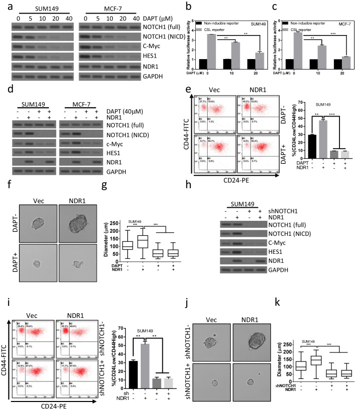
Fig. 4.
Activation of Notch1 signaling pathway is essential for NDR1 enhanced cancer stem cell properties in breast cancer cells. a SUM149 or MCF-7 cells were treated with the indicated concentration of DAPT for 72 h. Cells were harvested and subjected to western blot analysis. b, c SUM149 or MCF-7 cells were treated with the indicated concentration of DAPT for 72 h. Cells were harvested and subjected to luciferase reporter assay. d NDR1 or control vector were expressed in SUM149 or MCF-7 cells. Cells were treated with the indicated concentration of DAPT for 72 h. Cells were harvested and subjected to western blot analysis. e–g NDR1 or control vector was expressed in SUM149 cells. Cells were treated with the indicated concentration of DAPT for the indicated time. Cells were harvested and subjected to CD24/44 analysis (72 h, e) and sphere-forming assay (7 days, f and g). The representative spheres were shown in f. The distribution of sphere diameters was shown in g. h NDR1 or control vector was expressed in SUM149 cells. Cells were infected with lentivirus expressed shNotch1 or control shRNA for the indicated time. Cells were harvested and subjected to western blot analysis. i-k NDR1 or control vector was expressed in SUM149 cells. Cells were infected with lentivirus expressed shNotch1 or control shRNA for the indicated time. Cells were harvested and subjected to CD24/44 analysis (72 h, i) and sphere-forming assay (7 days, j and k). The representative spheres were shown in j. The distribution of sphere diameters was shown in k. The bar represents mean ± SD of three independent experiments (*: p < 0.05, **: p < 0.01, ***: p < 0.001)

