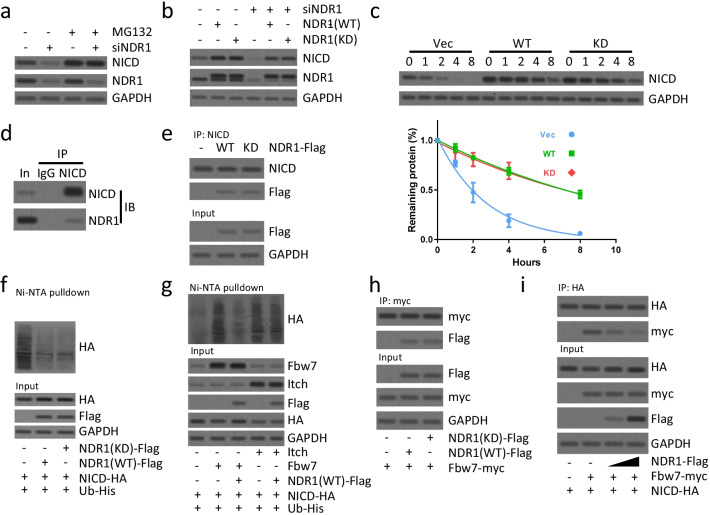
Fig. 5.
NDR1 suppresses FBW7 mediated degradation of NICD in breast cancer cells. a NDR1 siRNA or control siRNA were transfected in SUM149 cells for 48 h. Then, cells were treated with MG132 (10 μM) for 2 h. Cells were harvested and subjected to western blot analysis. b NDR1 siRNA or control siRNA were transfected in SUM149 cells for 48 h. Then, wild type or kinase-dead (K118A) NDR1 was overexpressed for 24 h. Cells were harvested and subjected to western blot analysis. c Wild type, kinase-dead NDR1 or control vector was overexpressed in SUM149 cells for 24 h. Cells were treated with CHX (50 µg/ml) for the indicated time. Cells were harvested and subjected to western blot analysis. The densitometric quantification of NICD normalized to GAPDH was plotted against various time points to determine the half-life of NICD. d The total protein of SUM149 cells was subjected to immunoprecipitation and western blot analysis using antibodies as indicated. e Wild type, kinase-dead NDR1 or control vector was overexpressed in SUM149 cells for 24 h. The total protein of SUM149 cells was subjected to immunoprecipitation and western blot analysis using antibodies as indicated. f, g SUM149 cells were transfected with indicated vectors for 24 h. Cells were subsequently treated with MG132 (10 μM) for 2 h prior to ubiquitination proteins were pulldown with Ni–NTA beads and followed by western blot analysis for indicated proteins. h, i SUM149 cells were transfected with indicated vectors for 24 h. The total protein of SUM149 cells was subjected to immunoprecipitation and western blot analysis using antibodies as indicated

