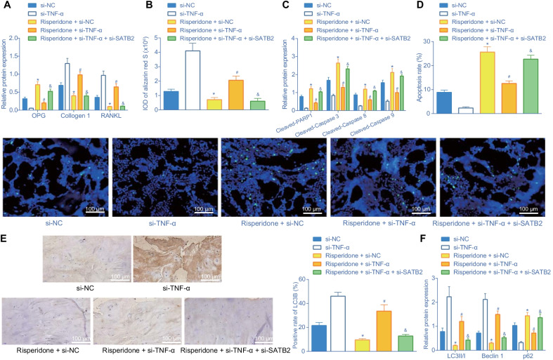
Fig. 8.
Risperidone represses differentiation and autophagy while enhancing apoptosis via the TNF-α/SATB2 axis in vivo. A, Western blot analysis of differentiation-related proteins (OPG, collagen I and RANKL) expression in femur tissues of mice. The band intensity was assessed. B, statistical results of alizarin red S staining in femur tissues of mice. C, Western blot analysis of apoptosis-related proteins (cleaved PARP1, cleaved caspase-3, cleaved caspase-8 and cleaved caspase-9) in femur tissues of mice. The protein band was assessed. D, apoptosis rate in femur tissues in mice assessed by TUNEL staining. E, representative images of femur tissues in mice stained by immunohistochemistry and statistical results of LC3B positive rate. F, Western blot analysis of autophagy-related proteins (LC3 II/I, Beclin1, and p62) in femur tissues of mice. The band intensity was quantified and analyzed. *p < 0.05, compared with mice introduced with lentivirus expressing si-NC. #p < 0.05, compared with mice introduced with Risperidone + lentivirus expressing si-NC. &p < 0.05, compared with mice introduced with Risperidone + lentivirus expressing si-TNF-α. The experiments are repeated 3 times n = 6

