Fig. 4.
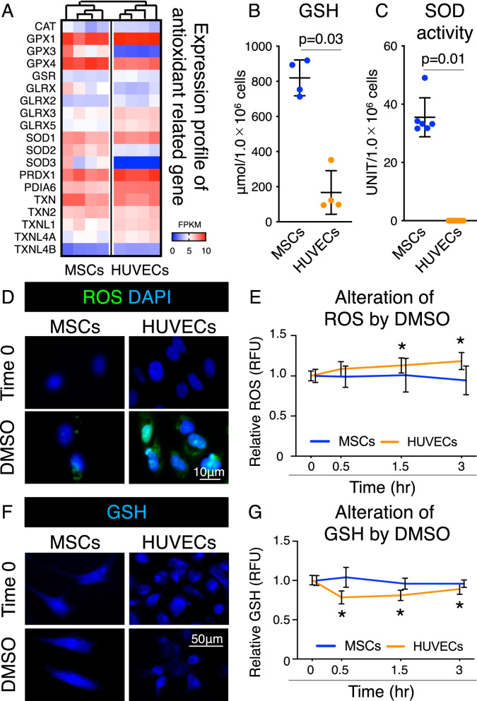
Antioxidant capacity of MSCs and HUVECs after DMSO supplementation. a Heatmap for expression profile of antioxidant-related genes. Cluster analyses are based on the expression profiles. FPKM: Fragments Per Kilobase of exon per Million mapped reads. b Amount of GSH in MSC and HUVEC lysates. Mean values and SD are shown (n = 4). The p value was calculated using the Mann–Whitney test. c Superoxide dismutase (SOD) activity in MSC and HUVEC lysates. Mean values and SD are shown (n = 6). The p value was calculated using the Mann–Whitney test. d Representative images of ROS (green) and cell nuclei stained with DAPI (blue). Data at time 0 and 3 h after supplementation with 5% DMSO are shown. e Alteration in ROS levels by 5% DMSO supplementation. Data are shown as relative fluorescence unit (RFU) values. *p < 0.05. The p value was calculated by two-way ANOVA with Dunnett’s multiple comparisons test. f Representative images of glutathione (GSH) (blue). Data at time 0 and 0.5 h after supplementation with 5% DMSO are shown. g Alterations in GSH levels by 5% DMSO supplementation. Data are shown as RFU values. *p < 0.05. The p value was calculated by two-way ANOVA with Dunnett’s multiple comparisons test

