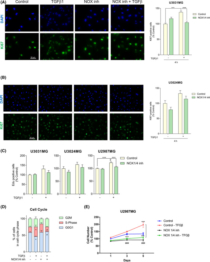
Fig. 3.

TGF‐β1 increases cell proliferation in a NOX4‐dependent manner. U3031MG (A) and U3024MG (B) cells were stimulated with TGFβ1 for 24h, in the presence or absence of the NOX1/4 inhibitor (GKT137831, 20 µm), and the following experiments were performed. (A, B) Left: Representative microscopic images of immunofluorescence of Ki67 (green) and DAPI (blue). Scale bar, 50 μm. Right: Quantification of Ki67 positive cells with respect to the control. Data represent the mean ± SEM (n = 2 for U3031MG and n = 3 for U3024MG independent experiment, 10 images per condition and experiments were quantified); statistics: two‐way ANOVA test, Sidak’s multiple comparison. (C) EdU staining was performed, data represent the mean ± SEM (n = 4 independent experiment with four biological replicates, nine images per condition and experiment were quantified); statistics: two‐way ANOVA test, Sidak’s multiple comparison. (D) Cell cycle was analysed by flow cytometry after staining the cells with propidium iodide, data represent the mean ± SEM (n = 4 independent experiment); statistics: two‐way ANOVA test, Dunnett’s multiple comparison. (E) Cell viability was assayed by MTS at the indicated times, data represent the mean ± SEM (n = 2 independent experiment with four biological replicates); statistics: two‐way ANOVA test, Tukey’s multiple comparison. (A–C, E) Statistical comparison indicates *P < 0.05, ***P < 0.001. (D) Statistical comparison indicates *P < 0.05, ***P < 0.001 vs Control; # P < 0.05, ### P < 0.001 calculated vs Control‐TGFβ.
