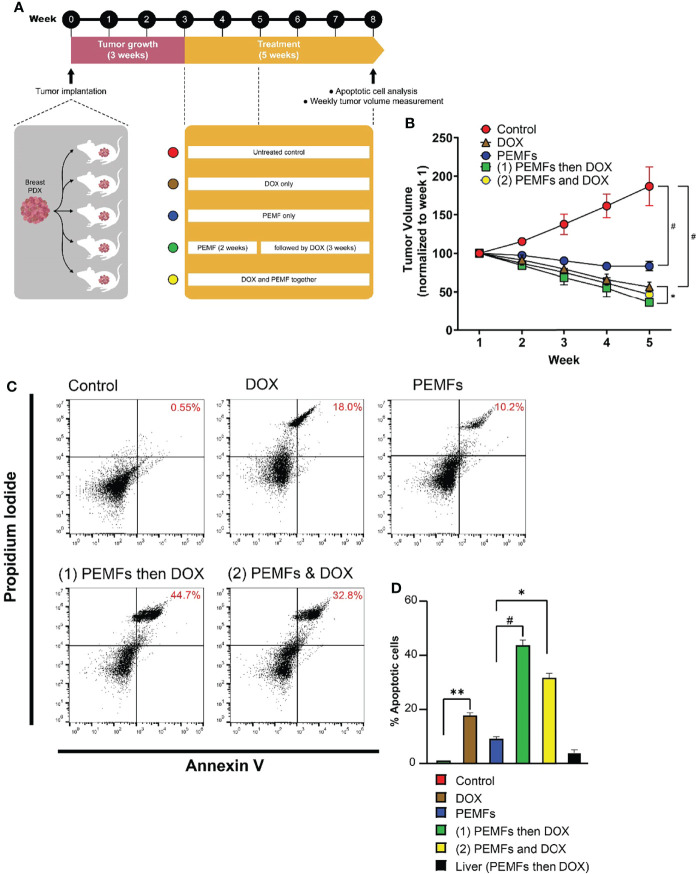
Figure 1.
PEMFs synergize with DOX to inhibit tumor growth in vivo. (A) Schematic of PEMF and DOX exposure regimes used on mice hosting patient-derived tumor xenografts. Implanted tumors were allowed to grow for 3 weeks before the initiation of DOX (20 mg/kg) and/or PEMF treatments. Tumor volumes were measured each week while apoptotic cell determination was performed at the end of the study. Each data point represents the mean values from 5 experimental runs derived from the tumors obtained from 5 patients, each of which was equally divided amongst the 5 treatment groups. (B) Changes in tumor volume (mm3) for 5 weeks. (C) Representative scatter dot-plots showing cell populations from dissociated tumors based on Annexin V and propidium iodide staining. (D) Quantification of apoptotic cell percentages obtained using flow cytometry. *p < 0.05, **p < 0.01, and # p < 0.0001. Error bars represent the standard error of the mean.

