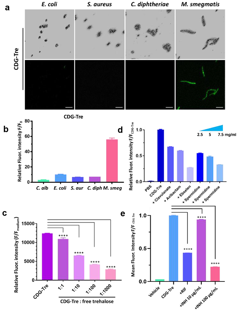
Figure 3.

Characterization of CDG-Tre labeling. (a) Confocal images (top row = bright field and bottom row = green fluorescence, Ex-490 nm/Em-520 nm) of freshly cultured E. coli (TOP10), S. aureus, C. diphtheriae and M. smegmatis incubated with 50 μM of CDG-Tre in PBS at 37 °C for 2 h. Scale bars indicate 5 μm. (b) Flow cytometry analysis of bacteria and fungi shown in (a). The unlabeled bacterial autofluorescence in PBS (F0) was utilized to normalize and calculate the relative fluorescence intensity. (c) Inhibitory study with increasing concentrations of free trehalose at 1, 10, 100, 1000-fold over CDG-Tre (100 μM) with M. smegmatis. (d) Inhibitory study with clavulanate (2 mg/ml), avibactam (2 mg/ml), ebselen (200 μg/ml) and spermidine (2.5, 5, 7.5 mg/ml) in the presence of 50 μM CDG-Tre with M. smegmatis. M. smegmatis treated with CDG-Tre exhibited a 60-fold increase of mean fluorescence intensity over PBS, which was arbitrarily set as 1 to normalize the test samples and show percentage inhibition. (e) M. smegmatis was treated with rifampicin (RIF, 0.2 μg/mL) or isoniazid (INH, 10 μg/mL or 100 μg/mL) for 3 h before incubation with CDG-Tre (100 μM) for 1 h at 37 °C. Mean fluorescent intensity of CDG-Tre was arbitrarily set as 1 to normalize other samples with drugs treatment and show percentage inhibition. All error bars represent standard deviation of three independent measurements. ****=P<0.0001
