
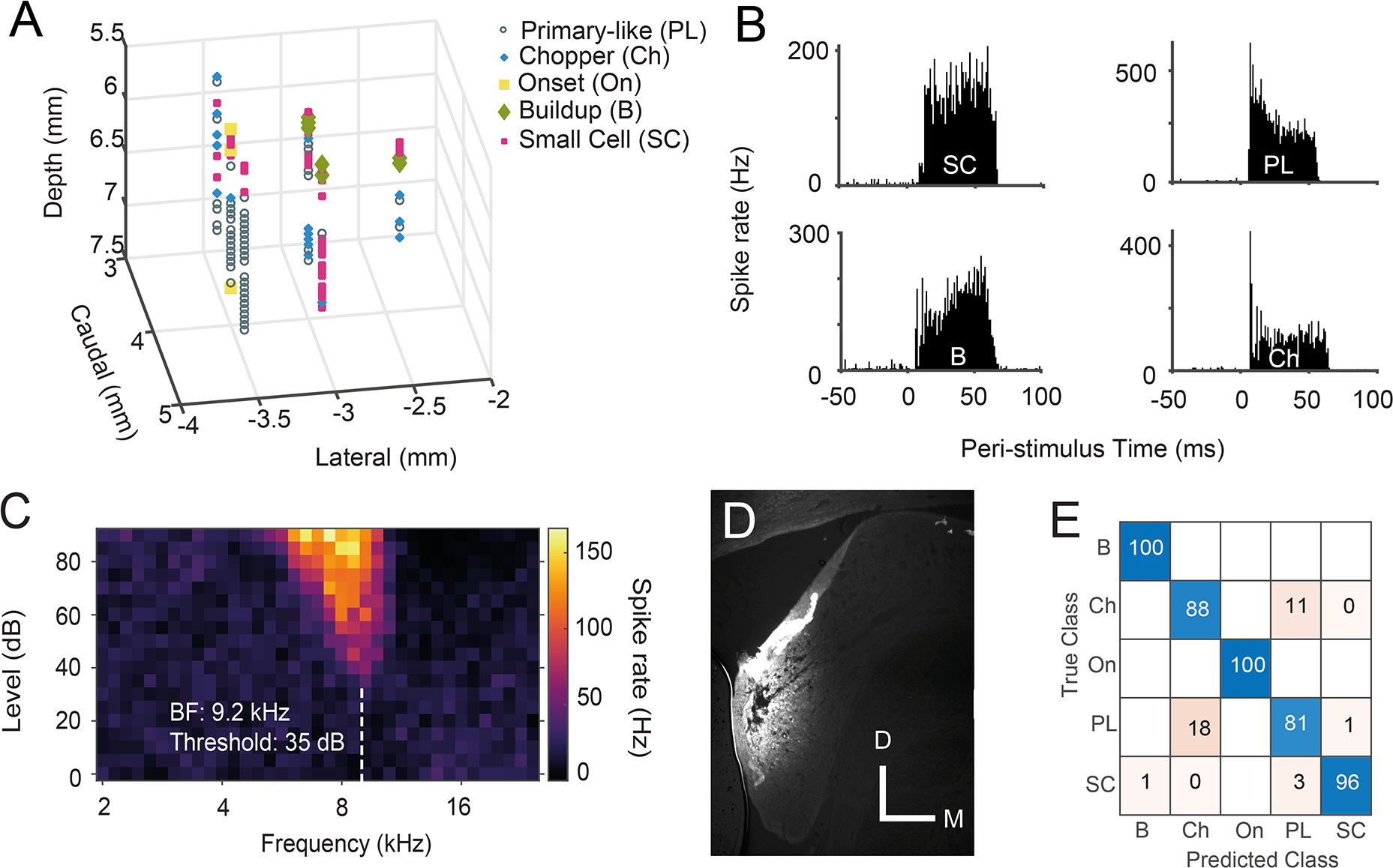
Fig 1: Validating recordings from CN small cells.
A) An example 3D plot of unit types within the left CN of one guinea pig. Cells exhibiting buildup PSTH shapes were found more medial and caudal, in the DCN. The VCN is more rostral and lateral, with the SCC units located between these two structures. B) Example PSTHs from 4 different CN cell types: small cell (SC) with flat, bimodal PSTH, primary-like (PL), buildup (B) and chopper (Ch). C) Receptive field of a CN small cell. D) Coronal section of the cochlear nucleus, containing a Fluorogold-marked electrode track in the SCC. (Bar = 0.5 mm) E) Confusion matrix of the machine learning model used to confirm recordings from discrete types of neurons. SC n = 303; PL n = 708; Ch n = 243; B n =185; On n =57.