FIGURE 1.
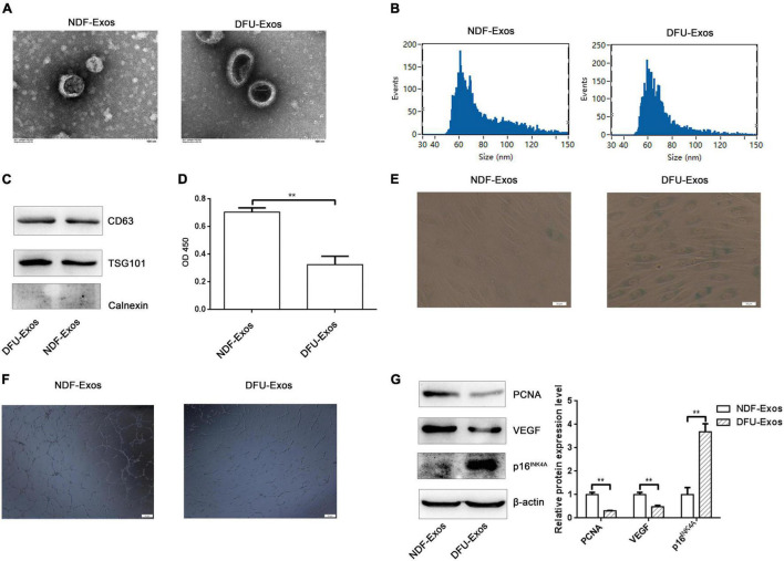
Exosomes secreted into the plasma of patients with DFU promoted cell senescence and inhibited tube formation in HUVECs. (A) TEM image showing the shape and sides of exosomes isolated from NDF-Exos and DFU-Exos. (B) NTA results showing the size of the exosomes. (C) Western blotting showing the expression of CD63, TSG101, and Calnexin in the exosomes. (D) Results of CCK-8 assay to detect the effects of NDF-Exos and DFU-Exos on cell viability. **P < 0.01 NDF-Exos vs. DFU-Exos. (E) β-gal stain showing β-galactosidase activity in HUVECs treated with NDF-Exos and DFU-Exos. (F) Tube formation in NDF-Exos and DFU-Exos groups. (G) Western blotting showing the expression of PCNA, VEGF, and p16INK4A in HUVECs incubated with exosomes. **P < 0.01 DFU-Exos vs. NDF-Exos.

