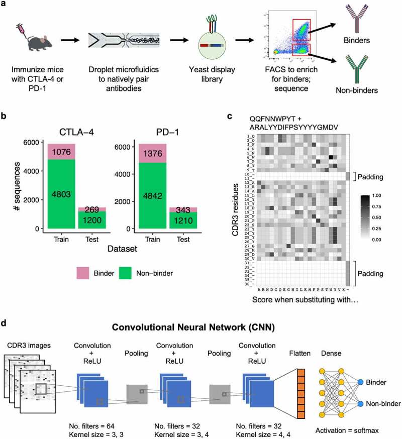
Figure 1.
Deep learning model for binder and non-binder classification. (a) Experimental workflow to generate antibody sequences. B cells from mice immunized with either CTLA-4 or PD-1 were encapsulated into microfluidics droplets to generate scFv libraries. The scFv libraries were expressed as yeast display, FACS sorted, and deep sequenced to generate binder and non-binder antibody sequences. (b) Number of CDR3K + CDR3H sequences in the training and testing datasets for CTLA-4 (left) and PD-1 (right). (c) A representative example of a CDR3K + CDR3H sequence encoded into a two-dimensional numerical matrix (image). The image displays BLOSUM substitution scores of the CDR3K + CDR3H residues (rows) when replaced with one of the 20 amino acids, gap, or X (columns). Both CDR3K and CDR3H were padded with “gaps” to ensure consistent dimension across images. (d) Convolutional neural network (CNN) model architecture for classifying binder and non-binder sequences. Two identical CNN models were built for PD-1 and CTLA-4 sequences, and they were trained separately.
(a) A flow diagram showing wet lab methods used to identify binder and non-binder antibody sequences, which were used to train the deep learning models used in this study. (b) A bar graph showing the numbers of binder and non-binder antibody sequences used for training and testing the deep learning models used in this study. Most antibody sequences were non-binders, and 80% of the antibody sequences were used for training versus 20% used for testing. (c) An example heatmap graph showing how antibody sequences are converted into images for processing by the deep learning model. Darker shades represent amino acids that are more likely to be substituted without a change in protein function. Only a small proportion of amino acids are indicated in dark shades in the figure. (d) An illustration showing convolutional neural network (CNN) model architecture for classifying binder and non-binder sequences.

