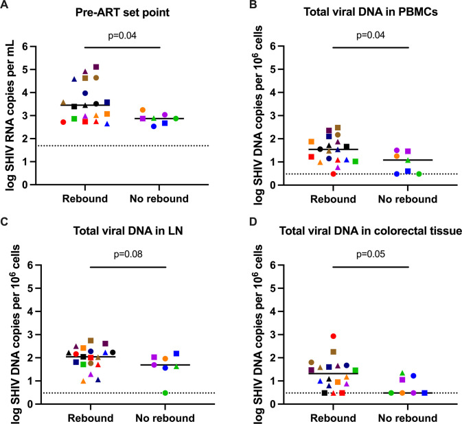
Fig 6. Analysis of viral rebound.
Comparison of (A) pre-ART set point viral load, (B) total viral DNA in PBMCs, (C) total viral DNA in lymph nodes and (D) total viral DNA in colorectal tissue at week 32 for animals that rebounded and animals that did not rebound following ART discontinuation. Dotted lines indicate limit of detection, values on the line were below limit of detection. Black lines indicate median values. Individual symbol and color coding as indicated in S1 Table.

